When the Connective Tissue Graft Was Too Good

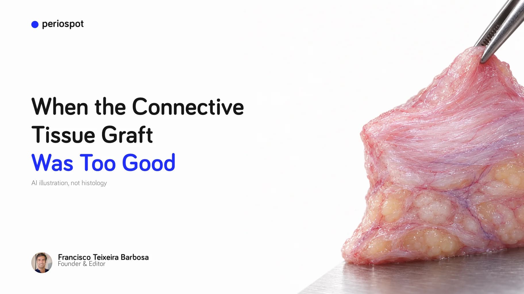
Table of Contents
There is a small, quiet celebration that happens in the surgery when you harvest a connective tissue graft and it comes out thick.
You hold it in the tissue forceps, dense and pale and generous, and a part of your brain lights up: this one is going to be beautiful. I have felt that celebration many times in my office in Barcelona. For years it was one of my favorite moments in all of surgery.
This article is about a case where that celebration betrayed me. It is about a maxillary central incisor, a graft that was, if anything, too good, and a complication that taught me more than any easy success ever did. And it starts with a question that sounds almost too simple to be interesting: when it comes to a connective tissue graft, is thicker always better?
A connective tissue graft is not just “connective tissue”
For most of my career I filed connective tissue grafts under one mental heading: “connective tissue.” Soft, pink-white, harvested from the palate, tucked under a flap. One material, one job.
That is not true, and the histology is quietly fascinating. A connective tissue graft is not one tissue. Depending on where you harvest it, how deep you go, and how you handle it, the same “graft” can be built from very different proportions of:
- Dense lamina propria, the collagen-rich, structural layer we actually want.
- Loose submucosa, softer and more compressible.
- Fatty and glandular tissue, the passenger nobody invited.
- Blood vessels and extracellular matrix, the living scaffolding.
The donor site changes the recipe. Human histology comparing the lateral palate with the maxillary tuberosity shows a real difference: tuberosity grafts tend to be dominated by dense lamina propria with very little loose submucosa, while lateral palatal grafts carry a much larger share of loose submucosa, fat and glandular tissue (Sanz-Martín and colleagues, 2019). In that study the tuberosity was roughly three-quarters lamina propria; the lateral palate closer to half, with about a quarter loose submucosa. More recent histology and gene-expression work points in the same direction.

One honest caveat, because it matters: “denser” here means a different composition, not a magically higher collagen content. Within the lamina propria itself, the collagen looked similar between the two sites. The tuberosity is not a better fabric. It just comes with less padding and less fat.


So the seductive idea, “thicker and denser is always better,” starts to wobble as soon as you look closely. A thick graft full of fat is not a good graft. A dense tuberosity graft is wonderful for volume, but it is not automatically the right answer for every problem. As I put it when I posted this idea online: the best graft is not always the densest graft. It is the one whose biology matches the recipient site's problem. Root coverage, implant volume, keratinized tissue, esthetics: different problems, different ideal grafts. Donor site selection is not only anatomy. It is biologic selection.

Hold that thought. Now let me show you the case where I forgot it.
The case: an old failure in the most unforgiving place

The patient came in unhappy with an old restoration in the upper front. A single maxillary central incisor, sitting on an implant that had already failed: peri-implantitis, poor esthetics, the works. The plan sounded clean on paper. Remove the failing implant, rebuild the site, place a new implant, and give it the soft tissue it deserved.
Clean on paper. The mouth had other plans.
Stage one: taking the implant out without wrecking the neighborhood
The first job was to remove the failing implant without destroying what little bone was left around it. The worst thing you can do in the esthetic zone is trephine an implant out and take the buccal plate with it.
Video 1. Removing the implant. Atraumatic counter-torque (reverse-torque, up to 200 N·cm) removal of the failing maxillary central-incisor implant, assisted by fine periotomes, with no bur cutting bone, followed by socket regeneration with the patient's own platelet fractions (PRP/PRF).
That part went well. The trap in this whole case was never the surgery I was worried about. It was the surgery I felt confident about.
Stage two: new implant, bone graft, and the graft I was proud of
A couple of months later, with the site healed, we went back in. A new implant into the central incisor position, submerged under a cover screw. Because the ridge was deficient on the facial, I did guided bone regeneration: particulate bone graft on the buccal, covered with a membrane, the classic sandwich. And then, to give the site the thickness and the pink resilience it would need long term, a connective tissue graft harvested from the palate.


Video 2. Placement, GBR and the connective tissue graft. Implant placement into the central-incisor site (after removal of the previously failed implant), with guided bone regeneration and a palatal connective tissue graft. In my own words in the original video description: “This case has a large story… A lot of mistakes but a lot of learning behind it.”
And here is where the celebration happened. The graft came out thick. Dense, generous, the kind you hold up and feel good about. I remember thinking, this site is going to have all the tissue it could ever want. I layered it in. I wanted maximum volume, maximum insurance.

When thick becomes a liability
Here is the biology I should have been thinking about while I admired my graft.
A free graft does not arrive with its own blood supply. For the first two to three days it survives by plasmatic diffusion, soaking nutrients from the recipient bed like a sponge, before new vessels grow in and reconnect over roughly the next two weeks. That diffusion only reaches so deep. Pile the tissue too thick, or lay it over a bed that cannot feed it, and the outer layer starves before the vessels arrive.
Now the honest nuance, and it is an important one. A connective tissue graft placed under a flap is usually forgiving about thickness, because it is fed from two sides: the bed underneath and the flap on top. That bilaminar blood supply is exactly why connective tissue grafts tolerate more bulk than a free gingival graft sitting exposed on periosteum. So thickness alone should not have killed this graft.
What killed part of it was the bed.

The thing under the tissue: the implant was exposed

The graft had been asked to survive over an implant that was exposed, with not enough bone covering it. No matter how good the tissue was, part of it was lying over a bare implant and a bone deficiency. Tissue cannot revascularize through titanium. I had given the site a beautiful graft and asked it to do a job that was never soft tissue's job to begin with.
I had been trying to fix a bone problem with soft tissue. And you cannot.
Fixing the floor before you paint the walls
So I stopped chasing soft tissue and went back to the foundation. That meant a second bone regeneration, and this time a vertical one. The first bone graft had gone in at implant placement, as part of the sandwich. Now, after the necrosis and the exposure, the site needed more than that: vertical bone augmentation over the exposed implant, particulate graft protected by a fixed, reinforced membrane, screwed down to hold the space and keep the graft still while it turned into bone. Rebuild the envelope first. Then, and only then, think about the soft tissue on top.


Let me be precise here, because the literature is precise. “You cannot fix bone with soft tissue” is true, but it has an edge to it. For a large hard-tissue deficiency, or an implant sitting in the wrong place, soft tissue will not save you: the bone and the implant position set the ceiling for what is possible. But for a small, contained buccal dehiscence of a few millimeters, soft-tissue augmentation alone can genuinely add thickness and hold up, and a recent randomized trial supports exactly that. The skill is not “always graft bone first.” The skill is reading how big the problem is, and matching the tool to it. Mine was not a small problem. I had treated it like one.
By the time the bone had regenerated and the soft tissue had settled over it, the site had changed completely. The tissue levels were finally acceptable. Only then, after the removal, two bone regenerations and the connective tissue graft, could we turn to the part patients think of first: the final restoration.


What I actually learned
- A thick graft is not a goal. It is a raw material. Ask what the site needs before you admire what you harvested. Volume, keratinized tissue, coverage and esthetics are different problems with different ideal grafts.
- Density and thickness are not the same thing. The donor site changes the recipe: tuberosity for dense, volume-stable tissue; lateral palate for more, but softer and fattier. Trim the fat. It does not revascularize, and nobody invited it.
- Under a flap, thickness is usually forgiving, until the bed cannot feed it. A graft over exposed titanium or bare, deficient bone is a graft over a surface that cannot give it blood. Respect the first three days.
- Soft tissue cannot rescue a hard-tissue problem. Read the size of the defect honestly. Small contained dehiscence: soft tissue can do it. Large deficiency or malposed implant: fix bone and position first, or accept the ceiling.
- Sequence is a decision, not an afterthought. The envelope, meaning bone and implant position, sets the ceiling. Build it before you decorate it.
The question I ask now
I still feel that little celebration when a graft comes out thick. I just do not trust it the way I used to. Now, when I hold up a dense, generous graft, a second voice asks the more useful question: not “isn't this a great graft?” but “is this the right graft, for this bed, for this problem, in this order?”
The best graft was never the thickest one. It was the one that matched the problem. This case cost me a complication to learn that. I am telling it here so that maybe it costs you only a few minutes of reading.
Selected references
- Sanz-Martín I, Rojo E, Maldonado E, Stroppa G, Nart J, Sanz M. Structural and histological differences between connective tissue grafts harvested from the lateral palatal mucosa or from the tuberosity area. Clin Oral Investig. 2019;23(2):957-964.
- Stuhr S, et al. Histological assessment and gene expression analysis of intra-oral soft tissue graft donor sites. J Clin Periodontol. 2023;50(10):1360-1370.
- García-Caballero L, et al. Histological and histomorphometric study of human palatal mucosa: implications for connective tissue graft harvesting. J Clin Periodontol. 2023;50(6):784-795.
- Bertl K, Pifl M, Hirtler L, et al. Relative composition of fibrous connective and fatty/glandular tissue in connective tissue grafts depends on the harvesting technique but not the donor site of the hard palate. J Periodontol. 2015;86(12):1331-1339.
- Müller HP, Schaller N, Eger T, Heinecke A. Thickness of masticatory mucosa. J Clin Periodontol. 2000;27(6):431-436.
- Studer SP, Allen EP, Rees TC, Kouba A. The thickness of masticatory mucosa in the human hard palate and tuberosity as potential donor sites for ridge augmentation procedures. J Periodontol. 1997;68(2):145-151.
- Sullivan HC, Atkins JH. Free autogenous gingival grafts. I. Principles of successful grafting. Periodontics. 1968;6(3):121-129.
- Guiha R, el Khodeiry S, Mota L, Caffesse R. Histological evaluation of healing and revascularization of the subepithelial connective tissue graft. J Periodontol. 2001;72(4):470-478.
- Zucchelli G, Tavelli L, Stefanini M, et al. Classification of facial peri-implant soft tissue dehiscence/deficiencies at single implant sites in the esthetic zone. J Periodontol. 2019;90(10):1116-1124.
- Ferrarotti F, Baima G, Romano F, Aimetti M. Peri-implant soft tissue increase at small buccal bone dehiscences with either a volume-stable collagen matrix or a connective tissue graft: a randomized controlled trial. Clin Oral Implants Res. 2025.
- Tavelli L, Barootchi S, Avila-Ortiz G, Urban IA, Giannobile WV, Wang HL. Peri-implant soft tissue phenotype modification and its impact on peri-implant health: a systematic review and network meta-analysis. J Periodontol. 2021;92(1):21-44.
- Thoma DS, Naenni N, Figuero E, et al. Effects of soft tissue augmentation procedures on peri-implant health or disease: a systematic review and meta-analysis. Clin Oral Implants Res. 2018;29(Suppl 15):32-49.
Check Your Knowledge
Comments
0 totalLoading comments...
Previous
Sinus Lift in Implant Dentistry: Lateral Window or Transcrestal Approach?
Next
25 years of implant follow-up: 94 percent survived, 14 percent met the health definition
Related Articles
Gum Disease and High Blood Pressure: What the Evidence Actually Shows
20 min read![Bundle Bone: The Complete Guide to Gap Management & Immediate Implants [2026 Update]](/_next/image?url=https%3A%2F%2Fajueupqlrodkhfgkegnx.supabase.co%2Fstorage%2Fv1%2Fobject%2Fpublic%2Fimages%2Fposts%2Fbundle-bone-the-complete-guide-to-gap-management-immediate-implants%2Fregenerated-1768916026108.jpeg&w=828&q=75)
Bundle Bone: The Complete Guide to Gap Management & Immediate Implants [2026 Update]
19 min read