Repair or regeneration
Rebuilding lost periodontal support
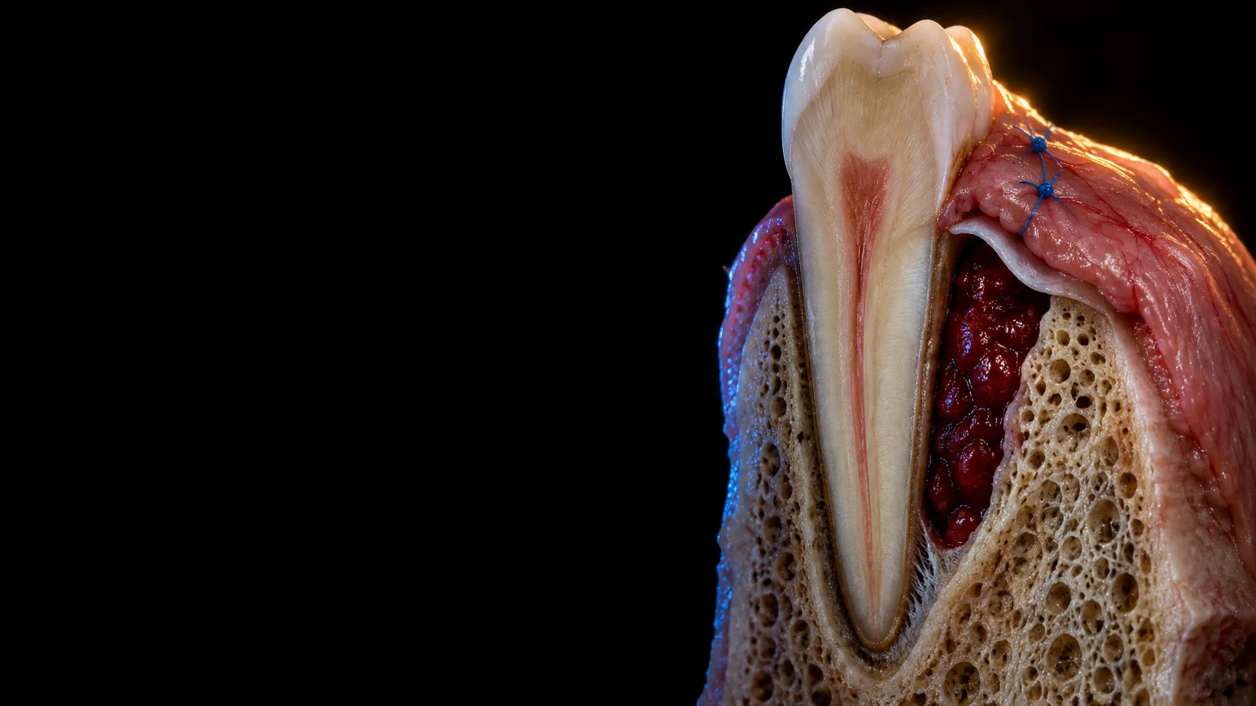
Rebuilding lost periodontal support requires fundamentally altering how periodontal wounds heal. Conventional therapy, such as scaling and root planing (SRP) or open flap debridement, predictably reduces probing depths and resolves soft tissue inflammation. However, classic block-section histology reveals that this healing occurs primarily through repair via a long junctional epithelium (LJE) rather than true structural restoration (Caton and Zander 1979; Caton et al. 1980).
Achieving true periodontal regeneration, defined as the re-establishment of new alveolar bone, a functional periodontal ligament (PDL) with inserting Sharpey's fibres, and new cementum on a previously diseased root surface, demands a targeted biological approach.
Section 1
The concept of compartmentalisation: Melcher's hypothesis
In 1976, A. H. Melcher proposed that the periodontium is divided into four distinct anatomical and cellular compartments:
- Gingival epithelium
- Gingival connective tissue
- Alveolar bone
- Periodontal ligament (PDL)
Cell competition and healing outcomes
Melcher's hypothesis posits that for true regeneration to occur, each compartment of missing tissue must be colonised by cells native to that specific tissue type. The theory was simple: whichever cell repopulates the debrided root surface first determines the outcome.
When inappropriate cell types colonise the wound bed, the original anatomical form is lost, resulting in repair or destruction rather than true regeneration.
Section 2
Experimental validation: the landmark animal studies
From 1980 onwards, Thorkild Karring, Sture Nyman, Jan Lindhe and colleagues published a seminal series of animal experiments verifying Melcher's concepts and disproving the notion that diseased root surfaces inherently prevent new attachment.
In summary, periodontitis-affected teeth in dogs and monkeys were extracted, root planed and implanted strategically so that the root surfaces were against bone (Karring, Nyman et al. 1980) or gingival connective tissue (Nyman, Karring et al. 1980). The PDL was removed on the coronal portions of the roots and carefully preserved in the apical portion.
- Karring, Nyman et al. (1980): In the areas where the PDL had been removed (coronal portion), healing was by repair, mainly root resorption and ankylosis, with no new attachment. In the apical portion, where the PDL was preserved, a functionally oriented attachment apparatus was reformed.
- Nyman, Karring et al. (1980): In the PDL-deprived areas, those facing bone showed consistent root resorption and ankylosis, while those facing connective tissue displayed "adhesion" of collagen fibres oriented parallel to the root, with no cementum. In areas where the PDL was preserved, the retained ligament formed a fibrous reattachment to the adjacent tissues.
Biological takeaway: Neither bone-derived nor gingival connective tissue-derived cells possess the biological capacity to establish new cementum with inserted collagen fibres.
In 1982, using a monkey model, Nyman, Gottlow et al. positioned a Millipore membrane before repositioning the flap after removing the buccal bone and curetting the cementum, in order to prevent the gingival connective tissues from meeting the denuded root surface during healing. Histological sections demonstrated new cementum with inserting collagen fibres on the curetted root surfaces, though not consistently along the entire length of the root. Healing at the coronal part of the wound was frequently characterised by connective tissue adhesion without signs of cementum formation.
Nevertheless, this suggested that periodontal ligament cells possess the ability to re-establish connective tissue attachment, providing the basis for the idea that if PDL cells were given preference, regeneration of attachment may be possible.
First human proof of concept
Nyman, Lindhe et al. (1982) applied this model to a human mandibular incisor with advanced periodontitis. After open flap debridement, a Millipore filter was secured beneath the flaps to exclude gingival connective tissue and epithelium.
After 3 months, histology demonstrated that epithelium proliferated along the outer surface of the filter rather than the root face, and new cementum with inserting collagen fibres formed along the root face, extending coronally from the PDL base up to 5 mm coronal to the bone crest.
Excluding the epithelium is not enough
Subsequent work by Karring, Isidor et al. (1985) in animal models showed that keeping the epithelium out by submerging the roots was not enough on its own: new attachment formed only in the apical part of the root, about 1 mm on average, by coronal growth of PDL cells. Thus, guided tissue regeneration (GTR) must exclude both epithelium and gingival connective tissue, giving strict preference to progenitor cells originating from the PDL.
The Root Experiments Behind Periodontal Regeneration
Companion video for this article. Vertical 9:16, 2:04.
Section 3
The biological conditions of guided tissue regeneration, and why meeting them is a challenge
To allow selective cell repopulation and regeneration to occur, three conditions set out by Susin, Fiorini et al. (2015) are necessary.
Space provision and maintenance
PDL progenitor cells require physical volume to proliferate and time to migrate into the space. Hence a barrier membrane or graft material must be capable of creating and maintaining space. If a barrier membrane collapses under pressure from the overlying mucogingival flap, the space is lost, adversely affecting regenerative outcomes.
In wide non-contained defects (such as 1-wall or 2-wall intrabony defects and wide furcations), the overlying soft tissue flap naturally collapses under muscle tension and mechanical loading, obliterating the sub-membrane space required for slow-migrating osteogenic and PDL cells.
Wound stability
Immediately after surgical closure, fibrinogen converts to fibrin, forming a fragile clot interface between the root surface and the soft tissue flap. Motion from mastication, tongue movement, swallowing or tooth mobility can disrupt this delicate fibrin link. Early detachment of the clot from the root is thought to allow epithelial downgrowth, shifting the outcome towards LJE healing (Wikesjö et al. 1992). Halting LJE formation is thought to depend heavily on stabilising this root-clot interface.
Primary intention healing
Primary intention healing, through tension-free flap adaptation and primary closure, protects the maturing clot during healing and reduces the risk of early membrane exposure and contamination of the site with oral microflora. More bacterial contamination of the membrane has been linked to less attachment gain (Selvig et al. 1992).
However, achieving primary intention closure over biomaterials or barrier membranes is technically demanding.
Despite a sound biological rationale, translating the principles of GTR into consistent clinical success remains remarkably challenging.
Why Periodontal Regeneration Is Biologically Difficult
Companion video for this article. Vertical 9:16, 2:08.
Hence, surgical techniques have continuously evolved, specifically in flap design and suturing techniques, to overcome these clinical barriers and provide the conditions for guided tissue regeneration:
- Minimal interdental tissue resection and papilla preservation flaps: Standard sulcular incisions often result in interdental soft tissue necrosis or flap tension over the biomaterial. The development of papilla preservation techniques, ranging from the Modified Papilla Preservation Technique (MPPT) (Cortellini et al. 1995, 1996) and the Simplified Papilla Preservation Flap (SPPF) (Cortellini et al. 1999) to modern Minimally Invasive Surgical Techniques (MIST and M-MIST) (Cortellini and Tonetti 2007, 2009) and Single-Flap Approaches (SFA) (Trombelli et al. 2009), aims to preserve the interdental soft tissue, move the incision away from the defect and protect the blood supply over the underlying membrane.
- Advanced suturing techniques: Precise microsurgical suturing, using internal mattress sutures, modified vertical mattress sutures and anchored mattress loops, allows clinicians to closely approximate interdental papillae and close the primary wound without tension. These suture configurations are designed to move mechanical stress away from the wound margin and underlying clot, supporting the wound stability required for PDL cell repopulation (Cortellini et al. 1999).
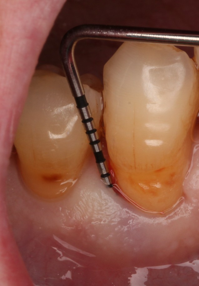
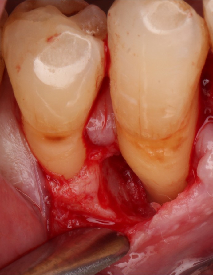
Clinical photographs: Jenny Wang.
A Stable Clot Starts With the Closure
Companion video for this article. Vertical 9:16, 2:02.
Section 4
The importance of case selection
If the goal is regeneration, every step must be planned carefully, starting with case assessment.
Regenerative outcomes are influenced by defect morphology. Wide, non-contained defects offer fewer bony walls to support the clot and supply cells, and wider defects gain less after GTR (Tonetti et al. 1993).
Not all defects or patients will be suitable for periodontal regenerative surgery. According to the EFP S3 level clinical practice guideline (Sanz et al. 2020), sites suitable for regenerative therapy are those where:
- Steps 1 (risk factor control) and 2 (non-surgical cause-related therapy) have been undertaken.
- The patient has adequate self-performed plaque control following steps 1 and 2 of therapy.
- The residual pocket is deep (PPD ≥ 6 mm) and associated with an intrabony defect 3 mm or deeper.
- There is a Class II furcation on a mandibular molar, or a buccal Class II furcation on a maxillary molar (a weaker recommendation).
The biological requirements of wound stability, space maintenance and primary closure apply whether using GTR membranes, bone grafts or enamel matrix derivative (EMD). When deep residual defects remain after re-evaluation, particularly in wide non-contained, complex or aesthetically sensitive sites, general practitioners should consider referring to a specialist trained in microsurgical regenerative protocols.
Jenny Wang
Specialist PeriodontistSpecialist periodontist with expertise in periodontal therapy, regenerative surgery and implant dentistry. Her clinical and educational work promotes evidence-based periodontology across the dental community.
Also by Jenny Wang: Why Does a Periodontal Pocket Become Shallower After Treatment?
Source register
References
- Caton JG, Zander HA. The attachment between tooth and gingival tissues after periodic root planing and soft tissue curettage. J Periodontol. 1979;50(9):462-466. PMID 114618 DOI
- Caton J, Nyman S, Zander H. Histometric evaluation of periodontal surgery. II. Connective tissue attachment levels after four regenerative procedures. J Clin Periodontol. 1980;7(3):224-231. PMID 7000854 DOI
- Cortellini P, Pini Prato G, Tonetti MS. The modified papilla preservation technique. A new surgical approach for interproximal regenerative procedures. J Periodontol. 1995;66(4):261-266. PMID 7782979 DOI
- Cortellini P, Pini Prato G, Tonetti MS. The modified papilla preservation technique with bioresorbable barrier membranes in the treatment of intrabony defects. Case reports. Int J Periodontics Restorative Dent. 1996;16(6):546-559. PMID 9242093
- Cortellini P, Pini Prato G, Tonetti MS. The simplified papilla preservation flap. A novel surgical approach for the management of soft tissues in regenerative procedures. Int J Periodontics Restorative Dent. 1999;19(6):589-599. PMID 10815597
- Cortellini P, Tonetti MS. A minimally invasive surgical technique with an enamel matrix derivative in the regenerative treatment of intra-bony defects: a novel approach to limit morbidity. J Clin Periodontol. 2007;34(1):87-93. PMID 17243998 DOI
- Cortellini P, Tonetti MS. Improved wound stability with a modified minimally invasive surgical technique in the regenerative treatment of isolated interdental intrabony defects. J Clin Periodontol. 2009;36(2):157-163. PMID 19207892 DOI
- Karring T, Nyman S, Lindhe J. Healing following implantation of periodontitis affected roots into bone tissue. J Clin Periodontol. 1980;7(2):96-105. PMID 6929795 DOI
- Karring T, Isidor F, Nyman S, Lindhe J. New attachment formation on teeth with a reduced but healthy periodontal ligament. J Clin Periodontol. 1985;12(1):51-60. PMID 3855871 DOI
- Melcher AH. On the repair potential of periodontal tissues. J Periodontol. 1976;47(5):256-260. PMID 775048 DOI
- Nyman S, Karring T, Lindhe J, Plantén S. Healing following implantation of periodontitis-affected roots into gingival connective tissue. J Clin Periodontol. 1980;7(5):394-401. PMID 6936413 DOI
- Nyman S, Gottlow J, Karring T, Lindhe J. The regenerative potential of the periodontal ligament. An experimental study in the monkey. J Clin Periodontol. 1982;9(3):257-265. PMID 6954167 DOI
- Nyman S, Lindhe J, Karring T, Rylander H. New attachment following surgical treatment of human periodontal disease. J Clin Periodontol. 1982;9(4):290-296. PMID 6964676 DOI
- Sanz M, Herrera D, Kebschull M, Chapple I, Jepsen S, Berglundh T, Sculean A, Tonetti MS; EFP Workshop Participants and Methodological Consultants. Treatment of stage I-III periodontitis: the EFP S3 level clinical practice guideline. J Clin Periodontol. 2020;47(Suppl 22):4-60. PMID 32383274 DOI
- Selvig KA, Kersten BG, Chamberlain AD, Wikesjö UM, Nilvéus RE. Regenerative surgery of intrabony periodontal defects using ePTFE barrier membranes: scanning electron microscopic evaluation of retrieved membranes versus clinical healing. J Periodontol. 1992;63(12):974-978. PMID 1474469 DOI
- Susin C, Fiorini T, Lee J, De Stefano JA, Dickinson DP, Wikesjö UM. Wound healing following surgical and regenerative periodontal therapy. Periodontol 2000. 2015;68(1):83-98. PMID 25867981 DOI
- Tonetti MS, Pini-Prato G, Cortellini P. Periodontal regeneration of human intrabony defects. IV. Determinants of healing response. J Periodontol. 1993;64(10):934-940. PMID 8277400 DOI
- Trombelli L, Farina R, Franceschetti G, Calura G. Single-flap approach with buccal access in periodontal reconstructive procedures. J Periodontol. 2009;80(2):353-360. PMID 19186978 DOI
- Wikesjö UM, Nilvéus RE, Selvig KA. Significance of early healing events on periodontal repair: a review. J Periodontol. 1992;63(3):158-165. PMID 1593409 DOI