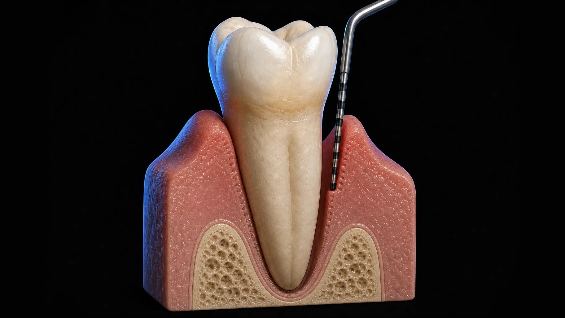
The clinical question
A wound that must reconnect four tissues to one root
Once biofilm and calculus have been disrupted, periodontal healing can begin. The periodontium is not a simple soft-tissue wound. Gingiva, periodontal ligament, alveolar bone and cementum must heal beside an avascular mineralised root surface, while the site remains exposed to oral microorganisms and mechanical loading.
A treated pocket can therefore become shallower, less inflamed and easier to maintain without rebuilding the original attachment apparatus. To understand why, we first need to look at how the periodontal wound heals.
Hours to days
The clot is the first scaffold
Within minutes of instrumentation or surgery, plasma proteins coat the wound surface and the coagulation cascade stabilises a fibrin clot against dentine or cementum. Platelets release mediators including platelet-derived growth factor, transforming growth factor beta and vascular endothelial growth factor.
Neutrophils arrive early to contain microbial challenge and clear debris. Monocytes follow and mature into macrophages, helping the wound move from inflammation toward reconstruction. Fibroblasts and endothelial cells enter the provisional matrix as granulation tissue becomes established.
Healing overlaps; it does not change phase at midnight
Select a process to see its illustration and explanation. The time windows are approximate and overlap, not fixed deadlines.
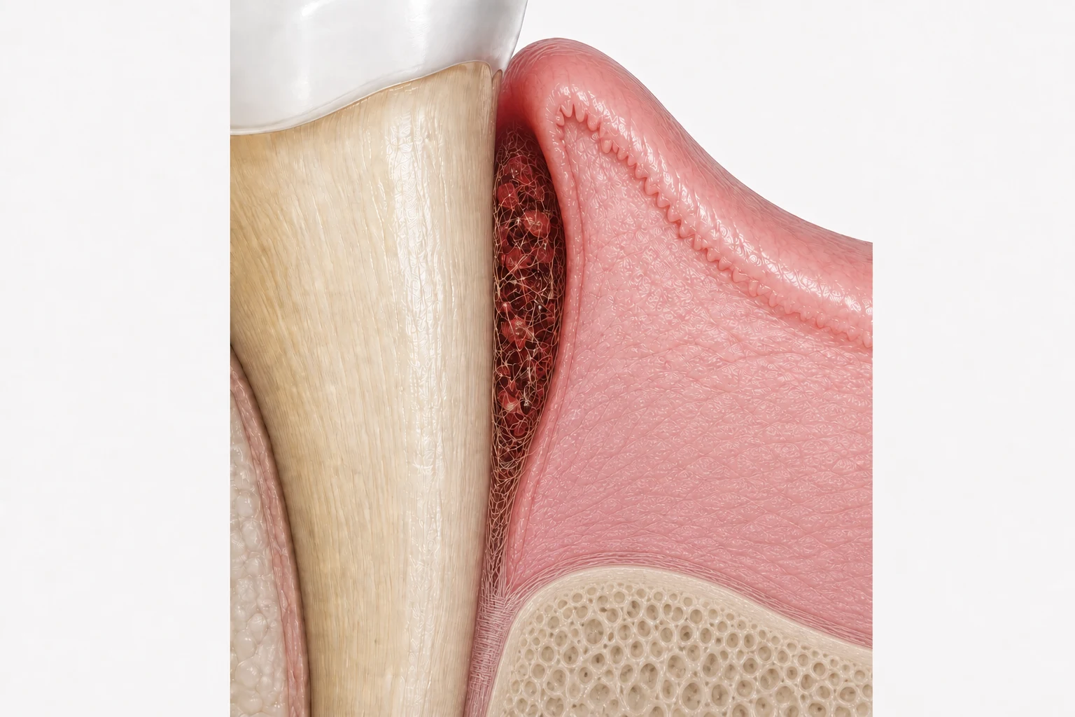
Clot
Fibrin stabilisation, platelet signalling, neutrophil influx and macrophage recruitment overlap as the wound is contained and cleared.
AI-generated teaching illustrations. These are process-focused views, not patient histology or a prediction of treatment outcome. Clot and inflammation share an early window; maturation and remodeling continue together. Bone regeneration is not implied.
Explore the clinical change
One pocket. Two changing signals.
Follow inflammation and tissue resistance through healing. Move the control to explore each stage.
Before treatment
An inflamed pocket wall is swollen and offers less resistance to gentle probing.
Illustrative trends, not patient data. Curves have no numerical scale and do not predict timing, treatment success or regeneration. Based on the tissue-condition explanation in the article and its cited probing studies.
Who arrives at the root
The first cells to repopulate the root surface influence the outcome
In 1976, A. H. Melcher proposed that the cells repopulating a previously diseased root surface influence the character of the attachment that forms. Epithelial cells can move rapidly along the root surface, so healing commonly produces a long junctional epithelium.
The pocket may look healthy and probe less deeply even when the original cementum, periodontal ligament and alveolar bone have not all been reconstructed. True regeneration requires cells with regenerative potential to reach the correct place, in the correct sequence, while the wound remains stable and protected.
Which cells shape the attachment?
Choose a tissue or tap a numbered marker. Then zoom in to see where it sits and what it contributes to healing.
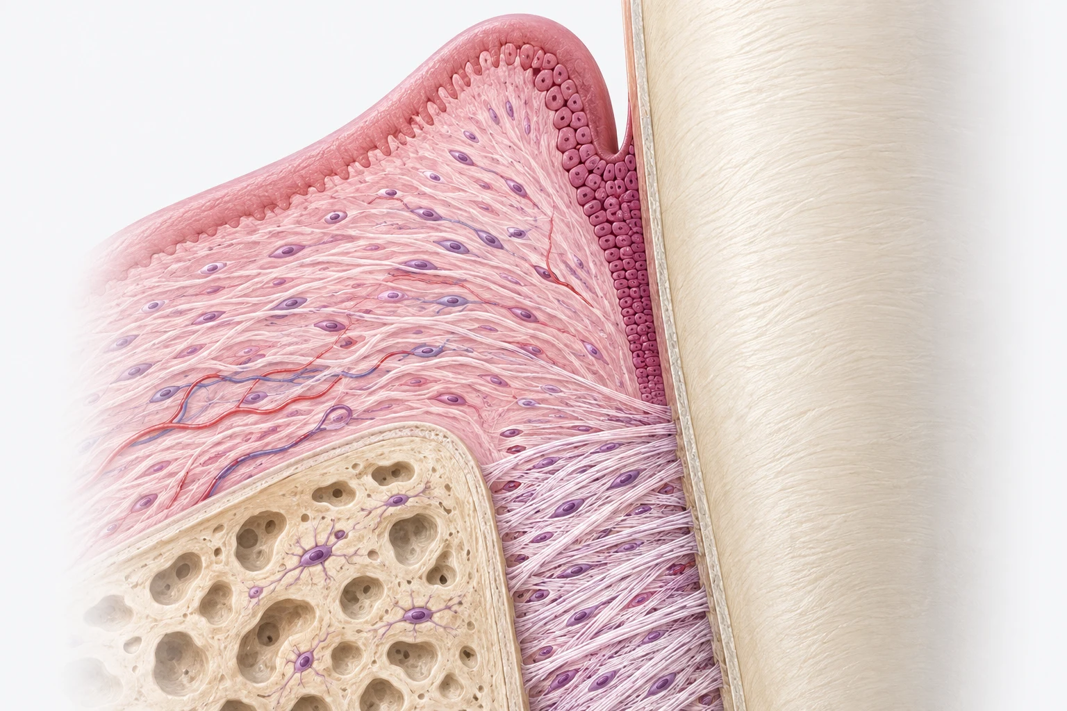
Long junctional epithelium
Epithelial cells can migrate rapidly along the instrumented root. The site may become clinically healthy and easier to maintain even when the original attachment apparatus has not been rebuilt.
What to look for: Follow the dark pink band next to the ivory root. This shows where the epithelial seal meets the tooth.
An epithelial seal is not the same as new cementum with inserting periodontal ligament fibres.
AI-generated anatomical teaching illustration. Cells and tissue proportions are simplified. This is a tissue explorer, not a model of migration speeds or a prediction of healing.
What the probe records
Why probing depth reduces after treatment
Before treatment, an inflamed pocket wall is swollen and mechanically fragile. The probe can pass farther through the inflamed tissue, sometimes beyond the apical end of the junctional epithelium. Probing depth is therefore influenced by tissue condition as well as anatomy.
After inflammation resolves, oedema decreases, the gingival margin may recede, collagen reorganises and the healed tissue offers more resistance to probing. A long junctional epithelial attachment can provide a clinically stable seal. Together, these changes can make the measured pocket shallower.
Reduced probing depth is a meaningful treatment outcome. It indicates a site that is less inflamed and generally easier to maintain, but it does not identify every tissue at the root surface or prove periodontal regeneration.
Why does the probe stop sooner?
Switch the illustration to compare the tissue wall and the position of the probe tip.
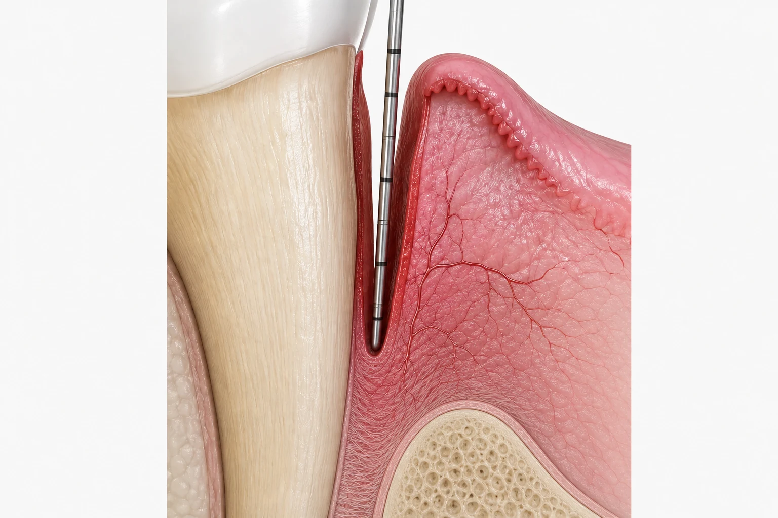
Less resistance, deeper penetration
Swollen, fragile tissue offers less resistance. A probe can penetrate farther into the inflamed pocket wall.
- Swollen gingival margin
- Inflamed soft-tissue wall
- Probe reaches farther apically
AI-generated teaching illustrations. Not to scale. Probe position also depends on force, angulation, tissue inflammation and local anatomy. Probing study.
Why Does the Pocket Become Shallower?
Companion video for this article. Vertical 9:16, 1:32.
Clinical appearance versus histology
A healthy result and a regenerated attachment are different endpoints
Clinical examination can show reduced probing depth, less bleeding and firmer tissue. Radiographs may also show increased density. These findings matter because they describe inflammation control, tissue stability and maintainability.
They cannot, on their own, reveal whether new cementum with inserting periodontal-ligament fibres has formed. That distinction requires histological evaluation, which is rarely available in routine patient care. The practical response is not to dismiss clinical improvement, but to describe the endpoint accurately.
Clinical meaning
Choose the endpoint before choosing the technique
If the objective is inflammation control and pocket reduction, healing by a stable epithelial attachment may be a clinically successful repair. Conventional periodontal therapy has not failed simply because histological regeneration has not occurred.
If the objective is true regeneration, treatment must create and protect a wound environment that supports the coordinated formation of new cementum, periodontal ligament and alveolar bone. That is a separate biological and technical question, and it deserves its own article.
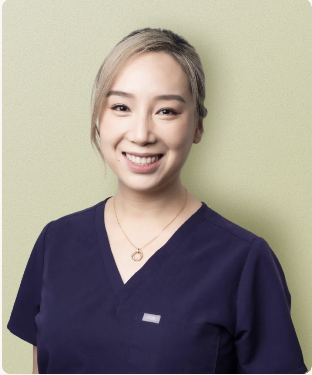
Jenny Wang
Specialist PeriodontistSpecialist periodontist with expertise in periodontal therapy, regenerative surgery and implant dentistry. Her clinical and educational work promotes evidence-based periodontology across the dental community.
Credentials and intended readers
Bachelor of Dental Science, Doctor of Clinical Dentistry (Periodontics), EFP Certified Specialist in Periodontology and Implant Dentistry
Intended readers: general dentists, dental students, hygienists and other dental professionals.
Source register
References
- Melcher AH. On the repair potential of periodontal tissues. Journal of Periodontology. 1976;47(5):256-260. PMID 775048 DOI
- Waerhaug J. Healing of the dento-epithelial junction following subgingival plaque control. II: As observed on extracted teeth. Journal of Periodontology. 1978;49(3):119-134. PMID 288899 DOI
- Caton JG, Zander HA. The attachment between tooth and gingival tissues after periodic root planing and soft tissue curettage. Journal of Periodontology. 1979;50(9):462-466. PMID 114618 DOI
- Listgarten MA, Rosenberg MM. Histological study of repair following new attachment procedures in human periodontal lesions. Journal of Periodontology. 1979;50(7):333-344. PMID 381631 DOI
- Armitage GC, Svanberg GK, Loe H. Microscopic evaluation of clinical measurements of connective tissue attachment levels. Journal of Clinical Periodontology. 1977;4(3):173-190. DOI
- Magnusson I, Listgarten MA. Histological evaluation of probing depth following periodontal treatment. Journal of Clinical Periodontology. 1980;7(1):26-31. PMID 6928855 DOI
- Grzesik WJ, Narayanan AS. Cementum and periodontal wound healing and regeneration. Critical Reviews in Oral Biology & Medicine. 2002;13(6):474-484. PMID 12499241 DOI
- Sculean A, Gruber R, Bosshardt DD. Soft tissue wound healing around teeth and dental implants. Journal of Clinical Periodontology. 2014;41 Suppl 15:S6-S22. DOI