Jaw bone density atlas · Text and sources clinically reviewed, September 2026
Inside the jaw, region by region: bone density in Hounsfield units.
A 3D teaching atlas of the maxilla and the mandible. Pick a region, see the CT density band one 2001 study proposed for it, then open a cross section that shows the cortical shell, the trabecular framework and the marrow space.
Runs in the browser on a laptop or a phone. No sign-in needed today.
What it shows
Three kinds of information, kept apart
The jaw map colours eight areas of the maxilla and mandible by CT density ranges reported in 2001, reproduced in 2012. Those are historical ranges for a region: they are not measurements of a patient, and the 3D model itself carries no density values. The cross sections are teaching models built in 3D by Periospot for five regions, drawn to illustrate the differences between sites reported in micro-CT studies rather than taken from those specimens. Blood supply and bone formation are explained in their own panels and drawn as optional layers, because how dense bone looks on a scan does not tell you how well it is supplied, and only loosely how one site will heal.
Reading the map
What the four density bands mean
Hounsfield units (HU) are the scale a CT scanner reports X-ray attenuation on. A higher number means more of the beam was absorbed.
These ranges are attributed to Norton and Gamble (2001) and are taken from the later reproduction by Reeves, Mah and McDavid (2012). Norton's original table has not been read directly. The grey values of an ordinary dental CBCT are not calibrated Hounsfield units, so a CBCT reading cannot be placed on this scale as it stands.
What you can do
Three ways to explore
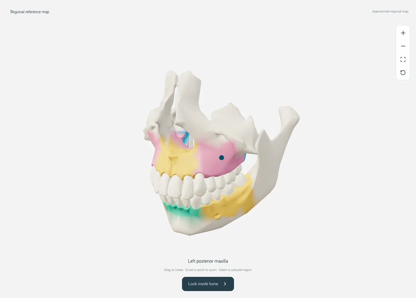
Jaw map
Rotate the maxilla and the mandible with 28 reference teeth. Select any of eight regions, show one jaw or both, hide the teeth, turn the regional colours off, and switch between front, side, top and three-quarter views.
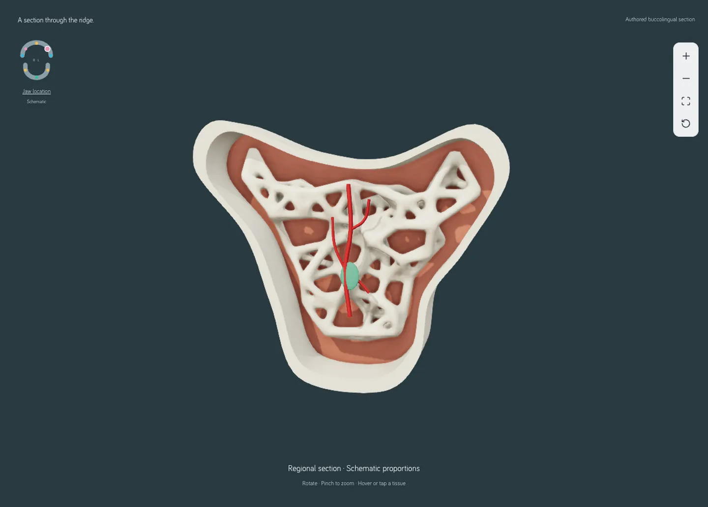
Inside bone
Open the cross section drawn for the selected region and tap the cut surface to name what you are looking at: cortical shell, trabecular bone or marrow. Marrow, illustrative vessels and bone-forming surfaces each switch on and off.
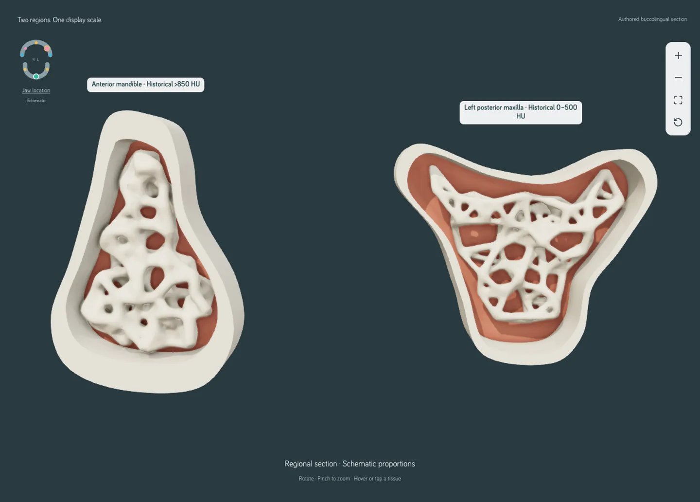
Compare
Put two regions side by side at the same display scale, for example the anterior mandible next to the posterior maxilla, and see how the shell and the framework differ between them.
The share button copies a link that reopens the same region, mode, layers and angle. Each region also carries short notes on architecture, mechanical support, blood supply, regeneration and imaging, and each note states what the evidence does and does not show.
Who opens it
How people use it
A student before an implant course
Open Compare with the anterior mandible and the posterior maxilla. The drawn sections illustrate the thinner trabeculae reported for the posterior maxilla (Kim et al. 2013), one reason that site is described as giving an implant less initial grip. How clinicians adapt the preparation is your course's job, not the atlas's.
A clinician talking to a patient
Show the back of the upper jaw next to the front of the lower jaw and explain why the bone there looks different on a scan, and why the plan may differ. The atlas does not tell you how that person's site will heal, and it says so on screen.
A teacher building a lecture
Set up a view, copy the share link into your notes, and take a screenshot for a slide. Credit the jaw surfaces to Z-Anatomy and BodyParts3D under CC BY-SA 4.0, and the cross sections to Periospot.
Expectations
What this is, and what it is not
You can expect
- Drawn teaching models for five regions, shown at one shared display scale.
- Regional notes that say where the evidence is thin, including a mouse study and a small graft study, both labelled as such.
- The same atlas on a phone, with Regions, Controls and Learn as drawers.
- A labelled still image if the device cannot run the 3D viewer.
The atlas does not provide
- A bone volume fraction, a density in HU, or any measurement in millimetres. The model's units are not calibrated.
- A prediction of implant stability, insertion torque, healing time or graft success.
- A drilling sequence or any treatment-planning output.
- A patient reconstruction, a micro-CT scan or histology.
- Tooth roots, the maxillary sinus or the mandibular canal inside the sections. They are not modelled, so their absence means nothing about real anatomy.
The text, the sources and the regional interpretation were clinically reviewed in September 2026; the 3D model geometry was not independently verified. Norton's original table has still not been read directly; the bands come from the Reeves 2012 reproduction, and the drawn sections remain teaching models.
Questions
Questions people ask about bone density
- What are Hounsfield units (HU)?
- Hounsfield units are the scale a CT scanner uses to report X-ray attenuation. A higher number means more of the beam was absorbed by the tissue. The Bone Quality Atlas shows the HU bands one 2001 study proposed for regions of the jaw; it does not measure HU on its own 3D model.
- What is the bone density of the anterior mandible compared with the posterior maxilla?
- In the regional CT density bands proposed by Norton and Gamble in 2001, as reproduced by Reeves, Mah and McDavid (2012): anterior mandible above 850 HU; posterior mandible and anterior maxilla 500 to 850 HU; posterior maxilla 0 to 500 HU; tuberosity below 0 HU. These are historical ranges for a region, not a value for a given patient or site, and the below 0 HU tuberosity figure is the one band no other published CT series we found reports, so treat it as Norton and Gamble's own observation. On the Hounsfield scale water reads 0 and air about -1000, so a value below 0 describes low attenuation, not absent or non-vital bone. Other CT series agree on the order but not the numbers: the anterior mandible averaged 945 HU in one series of 131 sites and 530 HU in another of 154 (Turkyilmaz et al. 2007; Fuh et al. 2010).
- Are CBCT grey values the same as Hounsfield units?
- No. In a study of ten cadaver mandibles (Parsa et al. 2012), cone beam CT grey values ran higher than medical CT Hounsfield units even though the two correlated closely. A CBCT reading cannot be placed on the HU scale as it stands.
- What is the Norton and Gamble bone classification?
- A 2001 study in Clinical Oral Implants Research (12(1):79-84) that proposed an objective bone density scale from CT Hounsfield values and reported a correlation between density, the subjective quality score and the region of the mouth. The four regional ranges shown on this page are the ones credited to it by Reeves et al. 2012; the original table has not been read directly. They also related the scale to the Lekholm and Zarb types, strongly for types 1 and 4, and proposed merging types 2 and 3 (as reported by Hiasa et al. 2011).
- How does this relate to the Lekholm and Zarb or Misch classifications?
- Lekholm and Zarb (1985) grade bone quality as types 1 to 4, assigned subjectively from the radiographic appearance of the cortex and the trabecular pattern, often with the drilling sensation at surgery, rather than from a number. Norton and Gamble related their CT values to those types: the correlation was strong for types 1 and 4, and they proposed merging types 2 and 3 (as reported by Hiasa et al. 2011). Misch's D1 to D4 scale is quoted with its own HU cut-offs (D1 above 1250 HU, D4 150 to 350 HU, as reproduced by Reeves et al. 2012), which differ from the regional bands on this page, so the colours cannot be read as D classes. Two of the atlas sources link the types to outcomes: Jaffin and Berman 1991 lost 35 of every 100 turned-surface implants placed in type IV bone against 3 of every 100 in types I to III, a historical figure rather than today's rate, and a 2017 meta-analysis of 94 studies (Chrcanovic, Albrektsson and Wennerberg) found type 4 bone still failed more often, while modern surfaces reduced the failure difference between type 1 and types 2 and 3, but not for type 4. A regional colour on the map is not a site's bone type.
- Does the atlas predict implant stability or tell me how to drill?
- No. It provides no bone volume fraction, no HU measurement of the model, no prediction of insertion torque, ISQ or healing, and no drilling sequence. The cross sections are drawn teaching models, not micro-CT, histology or a patient scan. A regional illustration cannot predict an individual site.
Evidence
Sources
Twelve published sources, jaw surfaces from Z-Anatomy and BodyParts3D (CC BY-SA 4.0), and cross sections drawn by Periospot. Each entry says what kind of study it is and what the atlas takes from it. Studies in mice or in small cohorts are marked, because they support a concept rather than a rule for every patient.
Regional CT density
- Norton MR, Gamble C. Bone classification: an objective scale of bone density using the computerized tomography scan. Clin Oral Implants Res. 2001;12(1):79-84. PMID 11168274 · doi:10.1034/j.1600-0501.2001.012001079.x
Human CT study. The regional density bands on the jaw map are attributed to this study. Its abstract reports that CT density correlated with the subjective quality score and with the region of the mouth; the full table of values has not been read directly, so the numbers shown here come from the 2012 reproduction below. - Reeves TE, Mah P, McDavid WD. Deriving Hounsfield units using grey levels in cone beam CT: a clinical application. Dentomaxillofac Radiol. 2012;41(6):500-8. PMID 22752324 · doi:10.1259/dmfr/31640433
Later reproduction of Norton regional bands. Its discussion section reproduces the four regional ranges credited to Norton and Gamble, which is where the exact numbers on this page come from. The study itself is about cone beam CT grey values. - Hiasa K, Abe Y, Okazaki Y, Nogami K, Mizumachi W, Akagawa Y. Preoperative computed tomography-derived bone densities in Hounsfield units at implant sites acquired primary stability. ISRN Dent. 2011;2011:678729. PMID 22203910 · doi:10.5402/2011/678729
Human CT study. Fifty-one patients and 104 implant sites that reached primary stability: the same regional order, anterior mandible highest and posterior maxilla lowest, with only four sites in the anterior mandible. It reprints Norton and Gamble's regional means and is the source for how they related their scale to the Lekholm and Zarb types: strong correlation for types 1 and 4, and a proposal to merge types 2 and 3.
Jaw bone microarchitecture
- Kim JE, Shin JM, Oh SO, Yi WJ, Heo MS, Lee SS, et al. The three-dimensional microstructure of trabecular bone: analysis of site-specific variation in the human jaw bone. Imaging Sci Dent. 2013;43(4):227-33. PMID 24380061 · doi:10.5624/isd.2013.43.4.227
Human jaw micro-CT study. Sixty-nine specimens showed site-related differences: outside the condyle, anterior mandibular trabeculae were the thickest and posterior maxillary trabeculae the thinnest. The drawn sections illustrate that reported difference. They are not built from these specimens. - Blok Y, Gravesteijn FA, van Ruijven LJ, Koolstra JH. Micro-architecture and mineralization of the human alveolar bone obtained with microCT. Arch Oral Biol. 2013;58(6):621-7. PMID 23123067 · doi:10.1016/j.archoralbio.2012.10.001
Human cadaver micro-CT. Ten cadavers: more bone volume and thicker trabeculae in the mandible, with no significant difference in tissue mineral density, and wide variation between and within people. - Ribeiro-Rotta RF, de Oliveira RC, Dias DR, Lindh C, Leles CR. Bone tissue microarchitectural characteristics at dental implant sites part 2: correlation with bone classification and primary stability. Clin Oral Implants Res. 2014;25(2):e47-53. PMID 23106552 · doi:10.1111/clr.12046
Human observational study. Thirty-two patients, relating microarchitecture to bone classification and insertion torque. It is the reason this atlas never predicts stability for a site: a regional illustration cannot do that.
Bone quality and implant outcomes
- Chrcanovic BR, Albrektsson T, Wennerberg A. Bone quality and quantity and dental implant failure: a systematic review and meta-analysis. Int J Prosthodont. 2017;30(3):219-37. PMID 28319206 · doi:10.11607/ijp.5142
Systematic review and meta-analysis. Ninety-four studies. Implants in Lekholm and Zarb type 4 bone failed more often than in types 1 to 3; modern surfaces narrowed the gap between type 1 and types 2 and 3 but not for type 4. A regional colour on this map is not a site's bone type, so this is context for the posterior maxilla note, not a prediction for a site. - Jaffin RA, Berman CL. The excessive loss of Branemark fixtures in type IV bone: a 5-year analysis. J Periodontol. 1991;62(1):2-4. PMID 2002427 · doi:10.1902/jop.1991.62.1.2
Retrospective implant series. One thousand and fifty-four turned-surface implants: 35 of every 100 placed in type IV bone were lost, against 3 of every 100 in types I to III. Older surfaces and a subjective bone type, so the number is history, not today's rate.
Imaging
- Parsa A, Ibrahim N, Hassan B, Motroni A, van der Stelt P, Wismeijer D. Reliability of voxel gray values in cone beam computed tomography for preoperative implant planning assessment. Int J Oral Maxillofac Implants. 2012;27(6):1438-42. PMID 23189294
Human cadaver imaging study. Ten cadaver mandibles: cone beam grey values ran higher than medical CT Hounsfield units even though the two correlated closely. This is why a CBCT reading cannot be placed on the scale above as it stands.
Biology and regeneration
- Kusumbe AP, Ramasamy SK, Adams RH. Coupling of angiogenesis and osteogenesis by a specific vessel subtype in bone. Nature. 2014;507(7492):323-8. Erratum in: Nature. 2014;513(7519):574. PMID 24646994 · doi:10.1038/nature13145
Mouse mechanism study. A mouse study linking a specific vessel type to bone formation. It supports the idea that vessels matter, and it is not a map of the vessels in a human jaw. - Wu V, Schulten EAJM, Helder MN, Ten Bruggenkate CM, Bravenboer N, Klein-Nulend J. Bone vitality and vascularization of mandibular and maxillary bone grafts in maxillary sinus floor elevation: a retrospective cohort study. Clin Implant Dent Relat Res. 2023;25(1):141-51. PMID 36214357 · doi:10.1111/cid.13142
Human graft cohort. Fifteen sinus-augmentation patients; tuberosity grafts showed more vitality and vascularisation than retromolar grafts at four months. It describes harvested graft tissue in one setting, not how every low-density site heals. - Wang HL, Boyapati L. “PASS” principles for predictable bone regeneration. Implant Dent. 2006;15(1):8-17. PMID 16569956 · doi:10.1097/01.id.0000204762.39826.0f
Regeneration framework. Primary closure, angiogenesis, space maintenance and stability, the framework behind the regeneration notes. Density on a scan is not one of the four.
Jaw surfaces and teeth come from the same open anatomy models used by the Anatomy Atlas. The cross sections were built in Blender by Periospot. Content lead: Francisco Teixeira Barbosa, DDS MSc.